The histopathology results were devastating. The mass was taken from the mid proximal (upper) left humerus (front upper bone). It was an oval mass measuring 2.5 cm in diameter. The dermis and subcutis were infiltrated by a “densely cellular pseudoencapsulated, moderately well-demarcated invasive neoplasm.” Cancer. The report continued, “Neoplastic cells are arranged in interlacing bundles, streams, herringbone patterns, and occasional whorls, with moderate amounts of basophillic, wispy stroma and collagenous stroma.” These are indicative of a sarcoma. “The neoplastic cells focally extend to one deep margin and abut the lateral surgical margin (separated from the surgical margin by only a few collagen fibers).” Bad news. What all this was saying was that he had a malignant subcutaneous fibrosarcoma that was incompletely excised. The margins weren’t clean. It was still there…growing, potentially spreading.
The tumor was classified as Grade II, intermediate. Again, we felt so guilty for not catching it earlier. Why hadn’t we seen it? Felt it? The pathology report said that cats with a grade II soft tissue sarcoma “had a median survival time of 514 days,” but the pathologist cautioned that “the grading scheme had not yet been validated by other studies.” He wrote that “feline fibrosarcomas in general tend to be highly locally invasive, with a metastatic potential that is low initially but appears to increase over time.” He continued stating, “In this case, neoplastic cells extend to deep margins, and there is high concern for local reoccurrence.” The report stated, that “aggressive surgical excision with wide margins appears to contribute to extended tumor-free interval and survival times in cats.” He recommended a consultation with a veterinary oncologist to discuss possible adjunctive therapies.
The report also said that there was “no overt histologic evidence to suggest this may be an injection site sarcoma (no peripheral aggregates of adjuvant-containing macrophages, no lymphoid follicle formation, no numerous multinucleated giant cells or marked pleomorphism).” The histopathology suggests it was a spontaneous fibrosarcoma. Bad luck. We checked the veterinary inoculation records. There is no record of him having a vaccine in that location. We had never given him the FeLV shot as he is indoors only but he had received the rabies vaccine as required by California law but not in that location. However, we adopted him at 6 mos. It is possible (likely) that he had vaccines prior to our adopting him. It is also possible a vet technician might have mixed up a left and right leg or that Castiel was being difficult at shot time, so the technician gave it elsewhere and didn’t note it in the paperwork. So many unknowns. He was also found by the rescue at a Maritime Academy, so he may have been exposed to something there or from his mum. Vaccine associated sarcomas have a worse prognosis than non-vaccine associated sarcomas, we’re told. We couldn’t be 100 percent sure which type it was, according to the vet. The vet said that it should be treated as though it was a vaccine induced sarcoma.
Every article I had been reading said the first surgery was the best chance to get all the tumor. We hadn’t gotten it. Would this lead to cancer seeding –spreading? These kinds of cancers are very difficult to get — they send out tentacles and they aren’t visible to the naked eye. Our vet had tried her very best but the monster was still there. And how long would it take to get an appointment with a veterinary oncologist? One place said they could see us in late December — too long. We called SAGE, they could get us in next week. Everything was taking so long but the report did say it was slow to metastasize so maybe there was hope but we worried, ” how long has it been there?”
The only encouraging news was that the X-rays looked clear and the blood work was mostly normal, only the globulin was slightly elevated. We made an appointment for Castiel with a veterinary oncologist and went home and collapsed.
Everything stopped. The wet laundry sat in the dryer growing mildew. The dishes sat in the sink. No one made dinner.
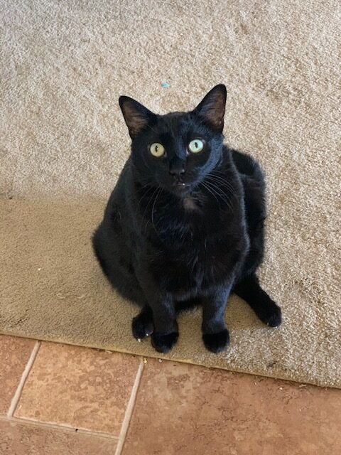
Awww buddy! He’s got a look like ‘come on guys, don’t worry, I’ve got this under control!’
Again, don’t beat yourself up. Our Jerry probably had osteosarcoma for months before we got the official diagnosis. Sometimes this is just how things happen. The important thing is you are taking action now.
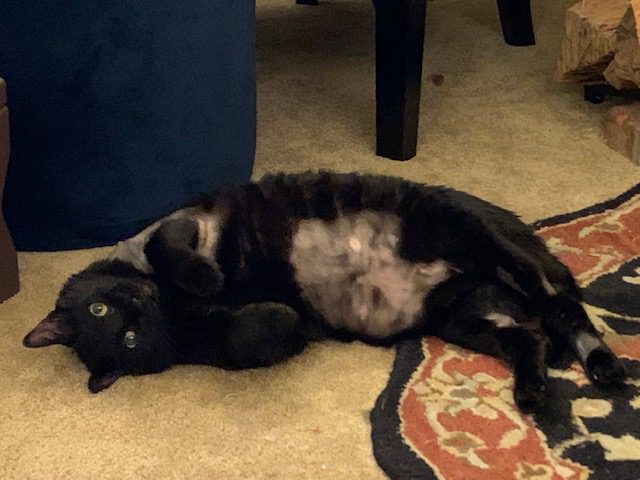
Thank you so much Jerry for all your support! Castiel is a very zen cat. We hope this surgery won’t change his personality. We hope he’ll still be our silly, goofy panther rolling about on jigsaw puzzles and batting his sister as she passes by.